APAP Model Introduction
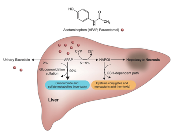
Yoon E , Babar A , Choudhary M , et al. [J]. Journal of Clinical & Translational Hepatology, 2016, 4(2):131-142.
APAP is taken up in the intestine within the first 2 hours after oral administration, metabolized in the liver via glucoronidation and sulfonation, and excreted in the urine. A small fraction (10-15%) is metabolized in hepatocytes by cytochrome P450 isomers to the alkylated, highly toxic metabolite N-acetyl-p-benzoquinoneimine (NAPQI). Antioxidant glutathione (GSH) converts NAPQI to the less harmful reduced form, which is then excreted via bile. When glutathione is depleted, increasing amounts of NAPQI bind to mitochondrial proteins and leading to hepatocyte necrosis.
In vivo efficacy of Celastrol in APAP Model

(A)Celastrol treatment alleviate increased ALT and AST enzyme activity induced by APAP.
Protective effects of Celastrol in APAP model
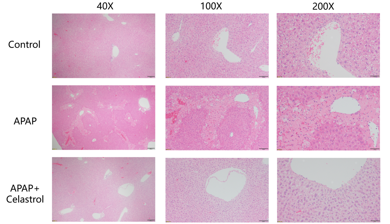
Protective effects of Celastrol in liver injury. Coagulative necrosis and bruising of cells in the marginal zone of the hepatic lobules were seen to varying degrees in the livers of the modeled animals, and the Celastrol treatment group showed significant improvement in the pathology.
Celastrol reduced apoptosis in APAP Model
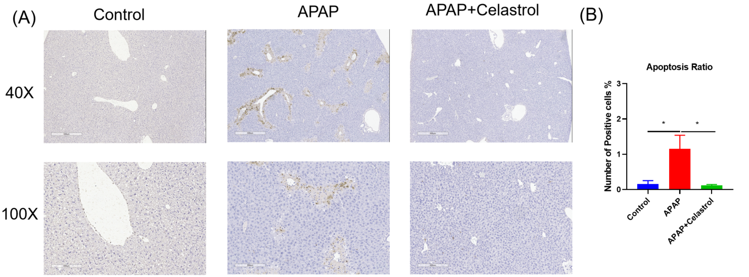
Celastrol reduced apoptosis in APAP induced liver injury. (A) Tunel staining showing apoptosis in hepatic cells. (B) Statistic data of Tunel positive staining.
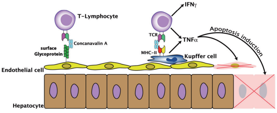
Heymann F, Hamesch K, Weiskirchen R, Tacke F.. Lab Anim. 2015 Apr;49(1 Suppl):12-20.
In vivo efficacy of Celastrol in ConA Model

Detection of liver injury-related factors in a ConA-guided NASH model after Celastrol intervention. (A) Celastrol treatment alleviate increased ALT and AST enzyme activity induced by ConA .
Effect of Celastrol on cytokines levels in ConA Model
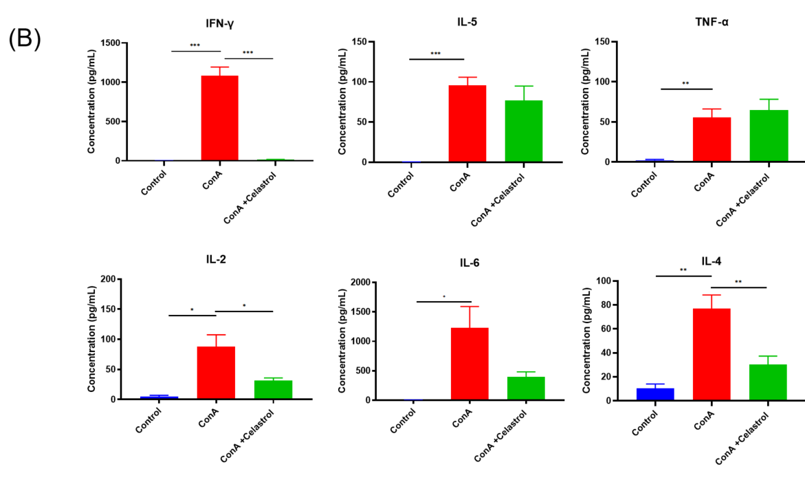
(B) Celastrol treatment alleviate increased cytokines levels induced by Con A. *:P<0.05; **:P<0.01; ***:P<0.001 compared with ConA group.
Protective effects of Celastrol in liver injury
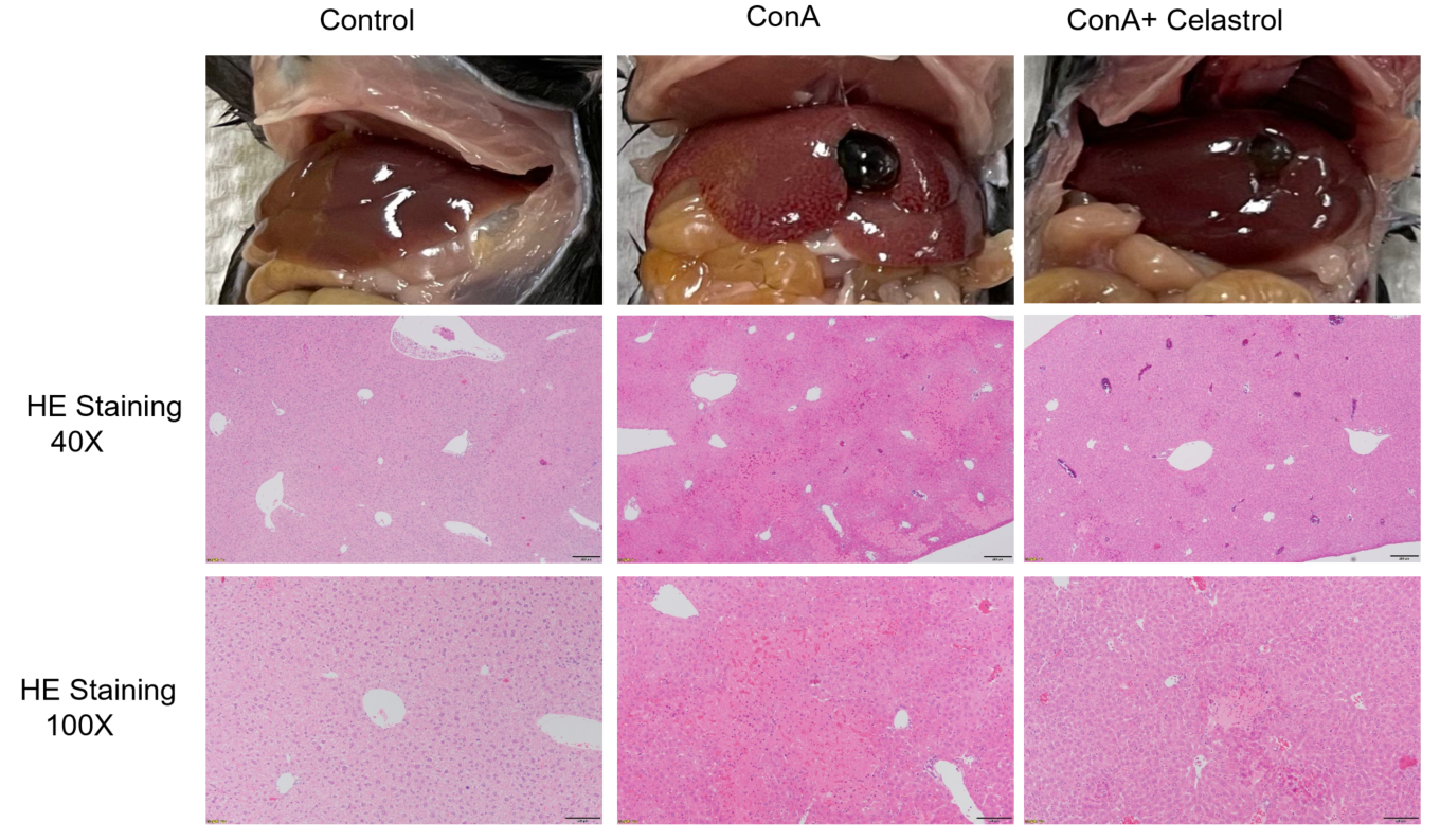
Analysis of H&E staining in the protective effect of Celastrol on ConA induced NASH mouse models.Coagulative necrosis and bruising of cells in the marginal zone of the hepatic lobules were seen to varying degrees in the livers of the modeled animals, and the Celastrol treatment group showed significant improvement in the pathology.

Pathological analysis in the protective effect of Celastrol on ConA induced NASH mouse models. (A) Tunel staining showing apoptosis in hepatic cells. (B) Statistic data of Tunel positive staining.


 +86-10-56967680
 info@bbctg.com.cn